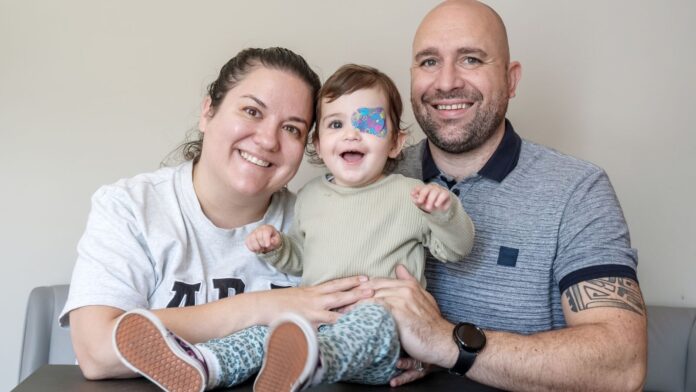
A toddler who had to have an eye removed after being diagnosed with a rare form of cancer has “adapted so well” to a prosthetic designed by doctors who relied solely on scans of her face.
Nuala Mulholland, now a 20-month-old toddler, was diagnosed when she was 10 months old, after her mother realised something was wrong as her eye appeared bloodshot.
It was initially thought Nuala, from Liverpool, had subconjunctival haemorrhage, a usually harmless condition where a tiny blood vessel breaks underneath the clear surface of the eye.
Read more on Sky News:
‘Very hot’ 30C days treble in UK, Met Office finds
What is the two-child benefit cap?
But her eye then started bulging out, prompting her mum, Megan, to take her to hospital.
Days later the family was informed she had a rare form of cancer which affects an average of only six people in England every year. The condition is known as alveolar soft part sarcoma.
“It was just horrendous,” Mrs Mulholland, 36, said.
“When I took her to A&E, I still didn’t think it was something as serious as cancer.”
The family was faced with the choice of removing the eye or going for radiotherapy, but were warned the latter could have a lifelong impact on Nuala because of her age.
Mrs Mulholland said: “Basically, for us, we felt like we had to make a good decision from two bad choices – radiotherapy or removing the eye.
“It felt like a rock and a hard place. We had to make the best decision for her.”
Nuala eventually had her eye removed and got the all-clear in January.
How did doctors design the prosthetic eye?
Doctors used a novel method to design and make Nuala’s prosthetic eye, which was less invasive than traditional methods, because of her age.
Patients who require this type of prosthetic usually face a lengthy process of having a wax mould taken of the eye socket, which is then turned into a silicone mould.
Nuala’s surgeon, Ankur Raj, a consultant in paediatric ophthalmology at Alder Hey, worked with the prosthetics team at Aintree University Hospital.
Mr Raj told PA: “You need to sit there for hours – you’re not going to get that with a one-year-old.”
In Nuala’s case, the team took a series of MRI scans, CT scans and photographs to help them reconstruct her face.
The MRI and CT images were used to shape the prosthetic, while photographs were used to match it to the position of the other eye. Colour matching to Nuala’s skin was carried out in person.
Another advantage to the prosthetic designed for Nuala is it wouldn’t have entailed the toddler being put to sleep, something her parents were relieved about as she had already been given anaesthetic about 15 times.
She had her first fitting in June, with doctors recommending she keeps it on a few hours each day so she gets used to it.
Mrs Mulholland said it is “amazing” what they have been able to achieve and praised the way her daughter has taken to her new prosthetic.
“Like everything she’s just adapted so well,” she said. “She takes a lot of it in her stride.
“She’s been really, really resilient,” she added.


